This website uses cookies so that we can provide you with the best user experience possible. Cookie information is stored in your browser and performs functions such as recognising you when you return to our website and helping our team to understand which sections of the website you find most interesting and useful.
Vyhledávání
Atomic force microscopy (AFM)
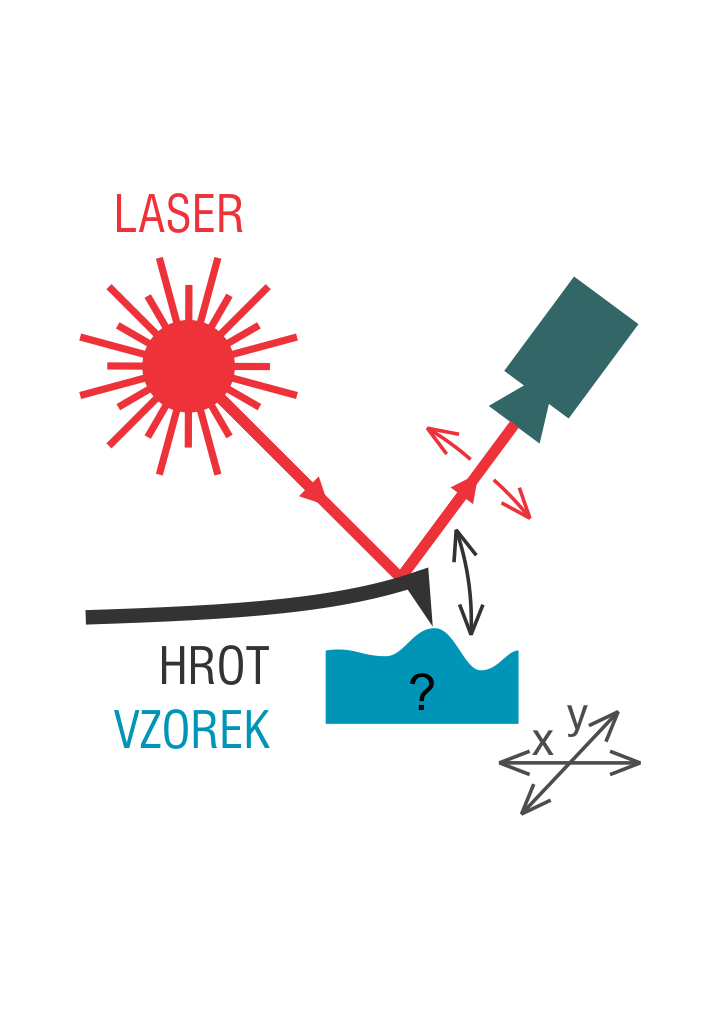
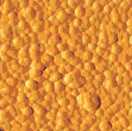
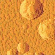
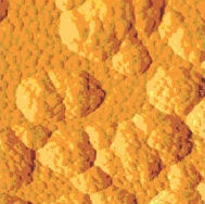
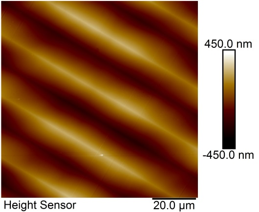
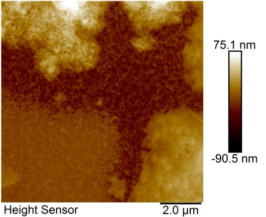
Atomic Force Microscopy(AFM) is a method for obtaining an image using a scanning probe (tip). The tip is placed on a cantilever and moved just over the surface of the studied sample, making a regular raster scan. Due to the irregularities of the surface, the cantilever tip bends slightly; this motion is detected by a laser. The dependence of the bending on tip position gives us surface topography. The image is built up point-by-point.
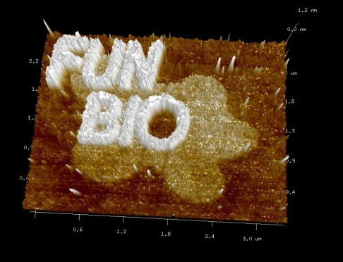
Measurements taken by the AFM method can be contact or non-contact. In the non-contact mode, the tip is oscillating over the surface up to the boundary of the resonance frequency, but close enough to the sample to maintain interatomic attraction between the tip and the sample (repulsive and attractive Van Der Waals forces). In the contact mode, the cantilever deflection is kept at a constant value, with the surface scanned by the feedback system. AFM can only display the surface of samples but its high-resolution is nearly comparable to that of the electron microscopy. Unlike electron microscopy, AFM provides a 3D image of the surface, while electron microscope can only provide 2D images.
The preparation of samples for the AFM method is very simple and requires no special conditioning such as polishing or electrodeposition. AFM can even work in liquid environments, which is an asset especially when studying biological preparations that can require that a scan is made in the preparations’ natural physiological environment so that their function or their response to changes in their environment can be observed.
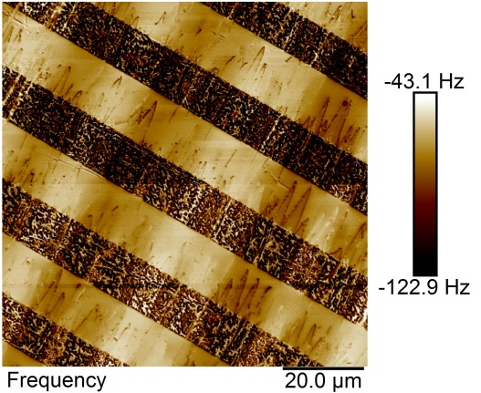
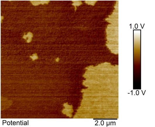
In addition to surface shape measurements, it is possible to perform more complex experiments. One of them is to measure local mechanical response based on the force-distance dependence, giving us information on material hardness or tip-surface adhesion. Such a measurement can be taken at every point, giving us the opportunity to obtain a map of mechanical properties of the sample’s whole surface. Using other, functional tips enables us to measure a great number of other parameters. The most common ones are magnetic domain distribution analysis, electrostatic field distribution analysis or surface potential analysis.
An obvious downside of AFM is a very limited range of measurements, the size of the resulting image (a maximum of hundreds of micrometers), and low imagining speed; the completion of an image takes up to several minutes. Also, it has a limited vertical range of measurements, typically amounting to the order of tens of micrometres, which is a limiting factor in terms of the sample’s maximum height. It is also the tip-sample proximity that may cause problems such as tips getting stuck or soiled. Sometimes also a sample damage can occur. Another problem is a non-zero tip width, which may cause an image distortion, compromising measurement accuracy.